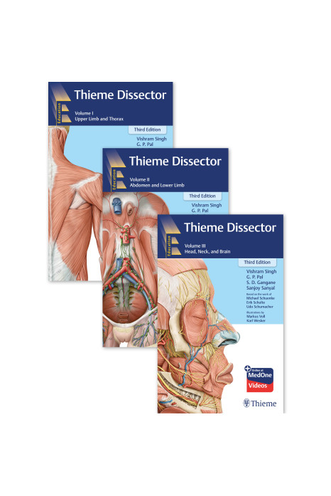
A Comprehensive Dissection Manual - Now in New Edition 2026 !!
Thoroughly revised for the modern learner, Thieme Dissector, Third Edition, is a richly illustrated and
detailed three-volume manual for guiding students and teachers in the dissection lab. It is enriched
with intricate illustrations of human anatomy created from the clinician’s perspective.
Carefully drawn
high-quality illustrations, QR code-based videos, and carefully researched and structured text guide the
students during dissection in a step-by-step manner.
Salient Features of the Third Edition
• Facilitates step-by-step introduction to dissection.
• QR code-based learning support: Provides access to more than 100 videos to facilitate learning and
enhance the scope of understanding the topic under discussion.
• Dissection screenshots: Presents most relevant and duly labelled clippings from the cadaveric
dissection videos for better insight into the steps of dissection.
• Radiographs: Includes radiographs to broaden the gamut of interpretation of the anatomy.
New to This Edition
• The content has been thoroughly revised and updated in accordance with the revised CBME
competencies (NMC curriculum 2024).
• Cadaver as the first teacher has been dedicatedly discussed separately in Volume I.
• Video list has been revised to make the book more relevant and student-friendly.
• New illustrations have been added to support improved anatomical understanding and accuracy.
• Computed tomography (CT) and magnetic resonance (MR) images of various regions have been
provided as an appendix to support clinical and radiological integration with anatomy.
ABOUT THE AUTHOR
Vishram Singh, MBBS, MS, PhD (hc), MICPS, FASI, FIMSA, Adjunct Professor, Department of
Anatomy, KMC, Manipal Academy of Higher Education, Mangalore, Karnataka, India; Editor-in-Chief,
Journal of the Anatomical Society of India; Member, Federative International Committee for Scientific
Publications (FICSP), International Federation of Association on Anatomists (IFAA), Geneva,
Switzerland.
G.P. Pal, MBBS, MS, FASI, FAMS, FNASc, FASc, Bhatnagar Laureate, Adjunct Professor, Department of
Anatomy, Index Medical College; Emeritus Professor, M.G.M. Medical College, Indore, Madhya
Pradesh, India.
S. D. Gangane, MBBS, MS, FAIMS, Former Professor and Head, Department of Anatomy and Genetic
Division, Grant Government Medical College and Sir J.J. Group of Hospitals, Mumbai, Maharashtra,
India.
Sanjoy Sanyal, MBBS, MS (Surgery), MSc (Royal College of Surgeons Edin.), ADPHA, Associate Dean
of Academic Affairs, Professor of Gross Anatomy, All Saints University School of Medicine,
Hillsborough Street, Roseau, Dominica.
Online at MedOne
Access your complimentary online version directly from https://medone.thieme.com by using the unique code in the front of this book.




-300x417.jpg)