Concise coverage of common temporal bone pathologies in a case-based format
Temporal Bone Imaging is a case-based review of the current techniques for imaging the various temporal bone pathologies frequently encountered in the clinical setting. Detailed discussion of anatomy provides essential background on the complex structure of the temporal bone, as well as the external auditory canal, middle ear and mastoid air cells, facial nerve, and inner ear. Chapters are divided into separate sections based on the anatomic location of the problem, with each chapter addressing a different disease entity.
Highlights:
- Each chapter features succinct descriptions of epidemiology, clinical features, pathology, treatment, and imaging findings for CT and MRI
- Bulleted lists of pearls highlight important imaging considerations
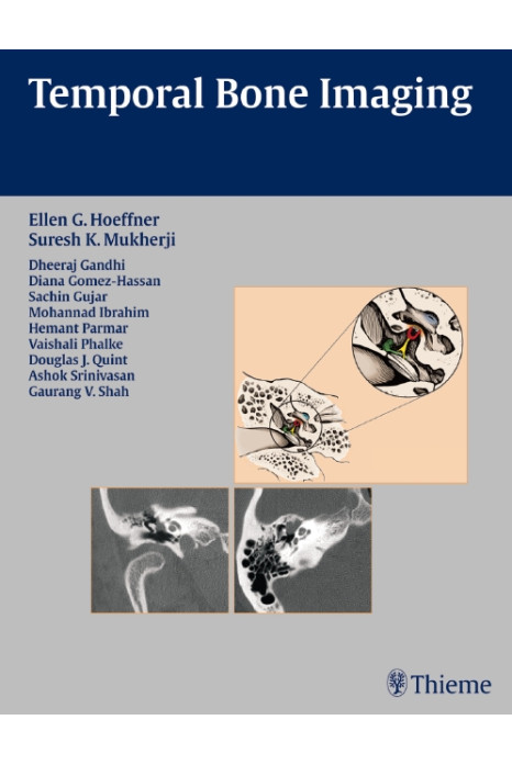
- More than 200 high-quality images demonstrate anatomy, pathologic concepts, as well as postoperative outcomes
This book will serve as a valuable reference and refresher for radiologists, neuroradiologists, otologists, and head and neck surgeons. Its concise, case-based presentation will help residents and fellows in radiology and otolaryngology-head and neck surgery prepare for board examinations.
Section I Anatomy
1 Temporal Bone
2 External Auditory Canal
3 Middle Ear and Mastoid Air Cells
4 Facial Nerve
5 Inner Ear
Section II External Auditory Canal
6 External Auditory Canal Atresia and Stenosis
7 External Otitis
8 Cholesteatoma of the External Auditory Canal
9 Exostoses
10 External Audiotry Canal Osteoma
11 Squamous Cell Carcinoma
12 Basal Cell Carcinoma
13 Melanoma
Section III Middle Ear and Mastoid
14 Ossicular Malformations
15 Congenital Cholesteatoma
16 Aberrant (Intratympanic) Internal Carotid Artery
17 Persistent Stapedial Artery
18 Dehiscent Jugular Bulb
19 Acute Ototis Media and Mastoiditis
20 Chronic Otitis Media
21 Acquired Cholesteatoma
22 Cholesterol Granuloma
23 Histiocytosis
24 Paraganglioma
25 Schwannoma
26 Hemangioma
27 Meningioma
28 Squamous Cell Carcinoma (Middle Ear)
29 Adenomatous Lesion
30 Adenoid Cystic Carcinoma
31 Rhabdomyosarcoma
32 Metastasis
Section IV Inner Ear and Petrous Bone
33 Cochlear Malformations
34 Semicircular Canal Dysplasias
35 Large Vestibular Aqueduct Syndrome
36 Internal Auditory Canal Stenosis/Atresia
37 Oval Window Aplasia/Hypoplasia
38 Cholesterol Granuloma of the Petrous Apex
39 Acute labyrinthitis
40 Labyrinthitis Ossifications
41 Petrous Apicitis
42 Vestibular Schwannoma
43 Meningioma
44 Congenital Cholesteatoma of the Petrous Apex
45 Epidermoid
46 Endolymphatic Sac Tumor
Section V Trauma
47 Transverse Temporal Bone Fractures
48 Longitudinal Temporal Bone Fractures
Section VI Postoperative Ear
49 Mastoidectomy
50 Ossicular Replacement Prostheses
51 Cochlear Implantation
Section VII Miscellaneous
52 Otosclerosis
53 Fibrous dysplasia
54 Paget Disease
55 Osteogenesis Imperfecta
56 Lymphoma
57 Superior Semicircular Canal Dehiscence