A practical approach to the early detection and management of breast cancer
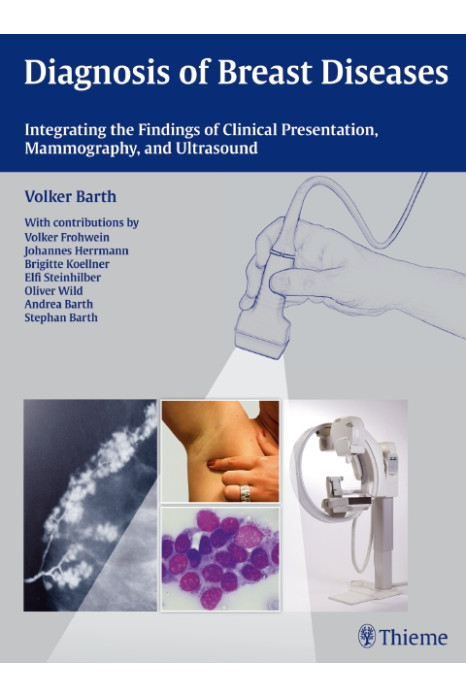
This atlas provides radiologists with essential information for the differential diagnosis of breast diseases on the basis of clinical presentation, mammography, and ultrasound. It begins with chapters on tumor biology, prognostic factors, and histology. The authors then provide a thorough evaluation of various methods for early detection and accurate diagnosis, including analog and digital mammography, ultrasound, MR imaging, PET/CT, and interventional procedures. They discuss in detail the strengths and limitations of each imaging modality, aspects of quality control, test intervals, peri- and postoperative management principles, and follow-up care.
Highlights:
- Presentation of difficult cases that effectively demonstrate the diagnostic hurdles and forensic pitfalls in breast diagnosis
- Special sections on breast cancer in men and young women, with discussion of women who are pregnant or lactating
- Color-coded practical tips and clinical notes for optimal comprehension of the material
- Extensive Q&A sections for self-testing in two major chapters
- More than 1,700 high-quality illustrations, including clinical color photographs, ultrasound images, and mammograms
1 Introduction
2 Aspects of Tumor Biology
Causes, Growth Factors, Endocrine and Exocrine Influences
Hormone Replacement Therapy in Menopause
Predisposing Genetic Factors
Case-Control Study on Sports and Breast Cancer
Summary
3 Prognostic Factors
Growth Rate, Tumor Size, Lymph Node Involvement, and Prognosis
4 Macroanatomy, Histology, Radiography, and Ultrasound
Anatomy of the Breast: Lobes, Lobules, and the Terminal Duct Lobular Unit (TDLU)
Terminal Duct Lobular Unit: Microradiography, Mammography, and Sonography
Conclusions
5 Early Detection and Appropriate Treatment
Diagnostic Options
Possibilities and Limitations of Complementary Investigations
Therapy and Perioperative Management
Postoperative Changes and Follow-Up
Answers
6 Summary
