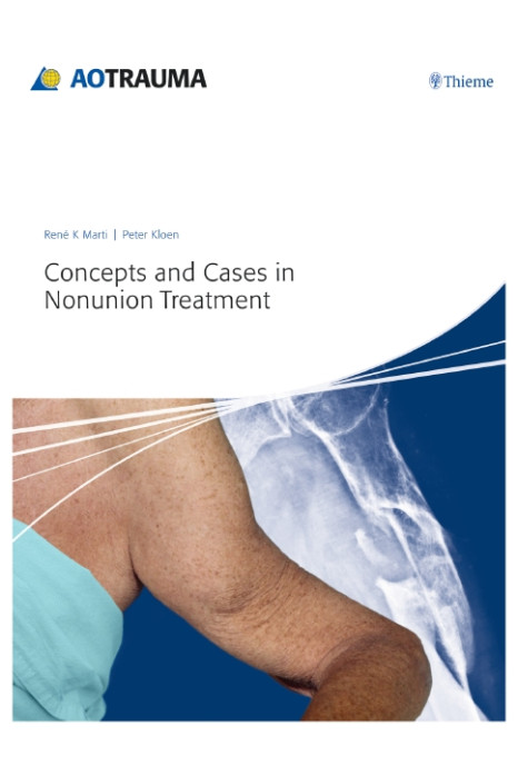
The gold standard for the treatment of nonunions was set by Weber and Cech in the early 1970s. With this new book the Editors René K Marti and Peter Kloen provide a comprehensive update on the state-of-the-art treatment of nonunions.
More than 130 case descriptions are included in the unique cases section; the core of this collection represents 40 years of René Marti's personal experience in nonunion treatment, demonstrating the principle "technique over technology". The editors have also carefully selected additional cases, contributed by several experts in nonunion treatment. Each case provides step-by-step descriptions of case history, preoperative planning, surgical approach, reduction, fixation, rehabilitation, and finally pitfalls and pearls. Hundreds of full-color pictures, precise illustrations, and x-rays demonstrate the significant steps in nonunion treatment.
In the principles preceding the case presentations relevant information on evolution, basic science aspects, nonoperative treatment, bone graft, as well as infected nonunions is provided. The guidelines and solutions presented for the management of nonunions support orthopedic and trauma surgeons worldwide.
1 Principles
1.1 Evolution of treatment of nonunions
1.2 Basic science aspects
1.2.1 Normal and impaired fracture healing
1.2.2 Pathogenesis and treatment of impaired fracture healing
1.2.3 Emerging treatments for fractures and nonunions—growth factors and beyond
1.2.4 Nonunion and the application of platelet-leukocyte gel (PLG) and bone morphogenetic protein (BMP)
1.3 Nonoperative treatment
1.3.1 Pulsed electromagnetic fields in the treatment of nonunions—the Dutch experience
1.3.2 Ultrasound in osteotomies and nonunions—basic research
1.4 Bone graft
1.4.1 The role of decortication in the treatment of nonunions
1.4.2 Autogenous bone grafting in the treatment of nonunions
1.4.3 Open cancellous bone graft
1.4.4 Bone-graft substitutes
1.4.5 Induced membranes—summary
1.5 Infected nonunions
2 Cases
2.1 Clavicle
2.2 Humerus, proximal
2.3 Humerus, shaft
2.4 Humerus, distal
2.5 Forearm
2.6 Pelvis/acetabulum
2.7 Femur, neck
2.8 Femur, proximal/intertrochanteric
2.9 Femur, proximal/subtrochanteric
2.10 Femur, shaft
2.11 Femur, distal
2.12 Tibia, proximal
2.13 Tibia, shaft
2.14 Tibia, distal/pilon
2.15 Ankle
2.16 Foot
