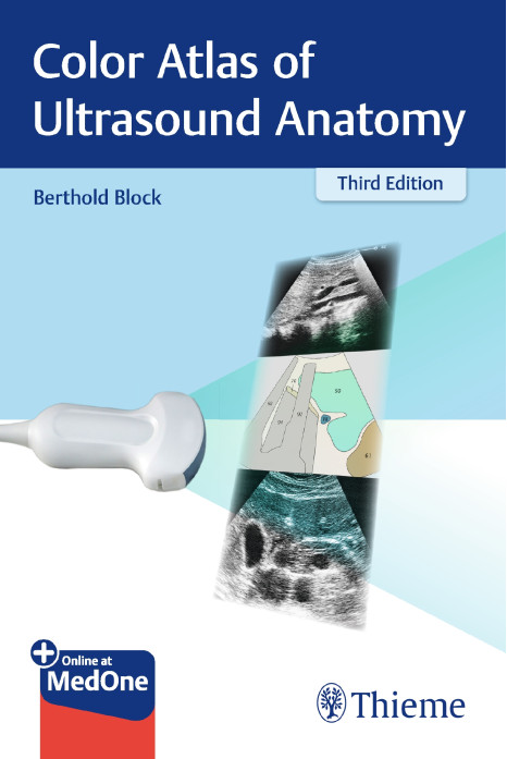
Beautifully illustrated with high-quality ultrasound images, an ideal beginner's guide; should be at hand in every ultrasound department.
Now in its third edition, the Color Atlas of Ultrasound Anatomy presents a comprehensive and systematic overview of normal sonographic anatomy of the abdominal and pelvic regions, essential for locating and recognizing the organs, anatomic landmarks, and topographic relationships. In its practical double-page format, ultrasound images and corresponding drawings are arranged by organs and scanning paths in more than 300 pairs, demonstrating probe positioning, the resulting sectional image, the anatomical structures, and the location of the scanning plane in the organ.
Special features:
- In gallbladder, spleen, and kidneys chapters, revised and expanded series of ultrasound images with corresponding drawings
- Now with coverage of transvaginal imaging of the uterus and ovaries and transrectal imaging of the prostate
- Offers guidance on scanning paths and standard sectional planes for abdominal scanning, with photos demonstrating probe placement on the body and drawings showing the organs that can be visualized
- Helps grasp the relation between three-dimensional organ systems and their two-dimensional representation in ultrasound imaging
- Front and back cover flaps displaying normal sonographic dimensions of organs for easy reference
Covering all relevant anatomic structures, important measurable parameters, and normal values, and including both transverse and longitudinal scans, this pocket-sized reference is an essential, high-yield learning tool for medical students, radiology residents, ultrasound technicians, and medical sonographers.
This book includes complimentary access to a digital copy on https://medone.thieme.com.
Standard Sectional Planes for Abdominal Scanning
1 Vessels
2 Liver
3 Gallbladder
4 Pancreas
5 Spleen
6 Kidneys
7 Adrenal Glands
8 Stomach
9 Bladder
10 Prostate
11 Uterus
12 Thyroid Gland


