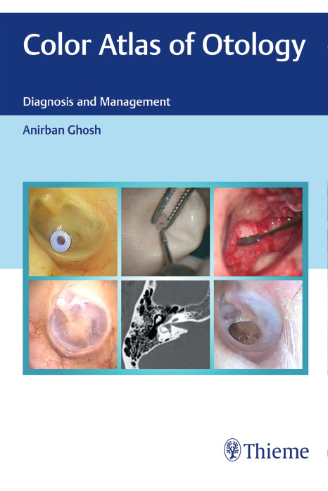
Color Atlas of Otology: Diagnosis and Management presents brilliant photographs of different otological disorders. The book has been divided into four sections covering all aspects of ear pathology, radiology, and surgical and postsurgical outcomes. All images are accompanied by a brief discussion about the disease and its management. High-resolution computed tomography (HRCT) of temporal bone has changed the scenario in cases of cholesteatoma and its complications, and a chapter has been added to give a detailed outlook on this. There are step-by-step detailed photographs of surgical procedures with explanations; the surgical steps are given in detail so that postgraduates and consultants can use them as a surgical road map to a particular disease.
Salient features:
Detailed color photographs of different ear pathologies.
CT scans of temporal bone for different complicated pathologies of ear diseases.
Step-by-step detailed images of different surgical procedures.
This book is a masterpiece of Otology. This book should be the companion of the residents from the very beginning. This can make the subject easier to understand and excel.
Dr Faizanul Haque
Royal College of Surgeons England , American College of surgeons
Posted on: 22 August 2024
Good book for PGs and Medical Practitioners
J A ROBERT
Sampoorna Hospital
Posted on: 22 August 2024
Excellent book. It is a must for all medical post graduate and consultant. Must for all medical library.
Complimentary copies are available to teaching faculty for review prior to course adoption. Please complete this form to request a complimentary copy. If you would like multiple copies, please have each person in your department fill out a request.
Have Questions?
Fill out the form given below and get all the answers to your questions.