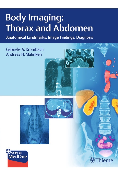
Body Imaging: Thorax and Abdomen reflects the realities of your everyday work: it describes the principal anatomic landmarks so that you can orient yourself in the chest and abdomen with speed and confidence, interpret the findings, and make a diagnosis.
Features:
- Description of key anatomic landmarks for rapid, confident orientation in the chest and abdomen
- Precise, step-by-step guide to making the diagnosis
- Key points summarized in boxes and tables
Comprehensive coverage
- All the modalities in one volume; no need for lengthy lookups in multiple books
- Focuses on the sectional modalities of CT and MRI, but includes plain radiographs and ultrasound as well
Answers to questions such as:
- Which modality is preferred?
- How are abnormalities recognized?
- How is the correct diagnosis derived?
- Differential diagnosis: What diseases are possible for any given set of symptoms and findings?
Richly illustrated
- More than 1500 superb images drawn from the latest generation of imaging technology, with explanatory diagrams showing details of anatomy and pathology
- Your radiology workstation in book form-structured, comparative, easy to use
Contents
- Part I: Chest
- 1. Mediastinum
- 2. Heart and Pericardium
- 3. Large Vessels
- 4. Lung and Pleura
- Part II: Abdomen
- 5. Liver
- 6. Gallbladder and Biliary Tract
- 7. Pancreas
- 8. Gastrointestinal Tract
- 9. Spleen and Lymphatic System
- 10. Adrenal Glands
- 11. Kidney and Urinary Tract
- 12. Female Pelvis
- 13. Male Pelvis
