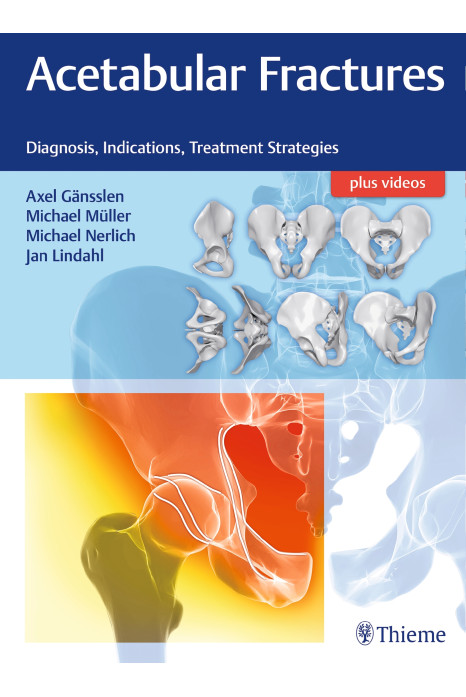
Enclosed within the deep and complex structures of the hip joint and the surroundings, acetabular fractures confront the orthopaedic surgeon with great challenges. A number of critical neurovascular structures in the vicinity are imperiled; the hip joint itself requires utmost care in surgery to preserve biomechanical stability over the long term and to postpone the development of posttraumatic osteoarthritis in the young to middle-aged patient collective.
It is the goal of this work to provide the surgeon with strategic tools to diagnose and evaluate the types of acetabular fractures to arrive at the optimal individual indication, thus taking a fracture-anatomy-guided approach to reduction and fixation.
Key Features:
- Eminently practical approach using more than 400 brilliant photographs, radiologic images, and drawings
- An emphasis on anatomical joint reconstruction to ensure the longest possible survival of the joint
- Discussion on age-specific problems and complications, such as osteoporosis, thromboembolism, and more
Acetabular Fractures will be welcomed by orthopaedic and trauma surgeons, as well as by residents and fellows, in these fields.
1 Surgical Anatomy
2 Biomechanics
3 Radiological Diagnostics
4 Classification of Acetabulum Fractures
5 Epidemiology
6 Indications and Planning
7 Approaches
8 Posterior Wall Fractures
9 Posterior Column Fractures
10 Associated Posterior Column and Posterior Wall Fractures
11 Anterior Wall Fractures
12 Anterior Column Fractures
13 Associated Anterior Column Plus Posterior Hemitransverse Fractures
14 Pure Transverse Fractures
15 Transverse plus Posterior Wall Fractures
16 T-Type Fractures
17 Both-Column Fractures
18 Fractures in the Elderly
19 Acetabular Fractures in Children
20 Heterotopic Ossifications
21 Thromboembolic Complications
22 Special Screws and Views
23 Outcome Scoring
24 Femoral Head Fractures: Pipkin IV
25 Periprosthetic Acetabular Fractures
